 There are 3 main causes of canine corneal lipid deposition.
1. Corneal lipid keratopathy or corneal lipidosis
2. Corneal lipid dystrophy
3. Corneal lipid degeneration
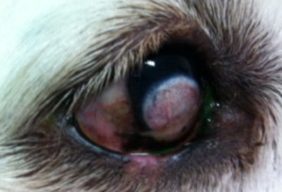
Corneal Lipid Keratopathy/Corneal lipidosis
This occurs when the corneal lipid deposition is secondary to metabolic diseases such as hypothyroidism, pancreatitis, hypercholesterolemia, diabetes, hypercalcemic conditions , Cushings disease, and plasma lipid elevations. Determining the underlying abnormality is essential in controlling this condition as is measuring fasting triglycerides and cholesterol levels. Keratectomy of the lipid plaques is contraindicated until the hyperlidaemic state is resolved as post-operative keratitis may lead to lipid depositing in the surgical site. In addition dietary management can help reduce the amount of lipid deposition.
Corneal lipid Dystrophy
Corneal lipid dystrophies are conditions that are typically non-inflammatory, non-painful and hereditary. They commonly occur bilaterally but at different rates. Depending upon the breed, the age of onset and location of the lipid in the cornea can vary. Whilst they may be slow to progress in some breeds like the Siberian Husky and Cocker Spaniel, they can be more rapid in other breeds such as the Airedale. Unfortunately the mode of inheritance is unknown for most breeds, but it is autosomal recessive in the Siberian Husky.
Corneal lipid Degeneration/ Calcareous Degeneration
Corneal inflammation, trauma and ageing can lead to lead to lipid deposition. These forms are associated with vascularisation of the cornea and can also contain calcium deposits. Differentiating lipid from calcium deposits is enhanced with slit-lamp biomicroscopy but often has a gritty texture to it. If calcium is identified than treatment with topical EDTA solution may help to dissolve the calcium. Whilst some forms may remain static other forms can be associated with ulceration and pain requiring medical management and sometimes surgery. It is not uncommon to see geriatric patients with corneal lipid degeneration leading to deep central ulceration requiring the placement of a conjunctival pedicle flap to prevent perforation and loss of the eye.


