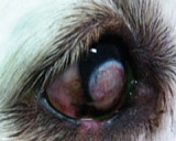
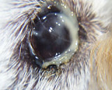
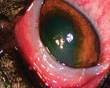
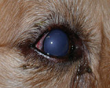
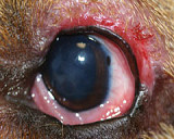
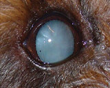
The normal IOP is 10 to 25 mmHg. Tonometry is the technique used to measure the IOP. Glaucoma can be defined as a raised intraocular pressure that affects the health of the optic nerve. Most dogs presenting with glaucoma have an IOP greater than 40 mmHg. It is very important that glaucoma is recognized very quickly to give the eye any chance of saving its vision.
When you should measure the IOP?
When you suspect glaucoma - the cardinal signs of glaucoma are a red eye, a blue eye and a mid-dilated non responsive pupil.
In any red eye - Uveitis and hyphaema can lead to glaucoma. Regular re-examination and tonometry allows you to chart the IOP, and diagnose glaucoma before vision is lost. Remember that you can use the IOP as a guide to your uveitis treatment. If the IOP was initially low eg 5 mmg, and has increased to 15 mmHg then your uveitis has been controlled. If the IOP in the uveitis treated eye goes from low to above 20 mmHg, secondary glaucoma is developing.
In all blue eyes - It could be glaucoma, or it could be an anterior lens luxation which usually ends up with secondary glaucoma, or uveitis in which the IOP should always be measured.
In any eye disease in pure bred dogs that may be prone to glaucoma - Check the breed predisposition list. Contact Animal Eye Care if you would like a list of glaucoma prone breeds.
Serial IOP measurements in dogs do not seem to have any benefit in predicting glaucoma in dogs. However serial IOP measurements in cats has been shown to help detect glaucoma in cats before it causes clinical signs and blindness.
How to measure the IOP
With any type of tonometry there can be potential errors, and as always the IOP readings should be correlated to the clinical signs. It is important to have a standardised technique to reduce measurement errors, this particularly applies to restraining the animal, avoid putting pressure on the eyeball and or eyelids, and or around the neck.
Local Anaesthetic is needed before using the Tonopen Avia, but is not required if using the the TonoVet. Aim to gently touch the tonometer in the centre of the cornea. Most electronic tonometers average 5 readings, repeat the procedure 3 times, and the actual IOP is closest to the lowest of these 3 readings.
Animal Eye Care uses both the TonoVet and the Tonopen Avia to measure the IOP. We are happy to provide advice on what tonometer to buy and how to measure the IOP.
